Lassonde professor explores optical imaging method for heart attack prevention
Tags:

Heart disease is a leading cause of death in Canada and the United States. Understanding the severity of the disease in patients is crucial for preventing complications like heart attacks – and researchers across the globe are racing toward solutions to avoid such devastating outcomes.
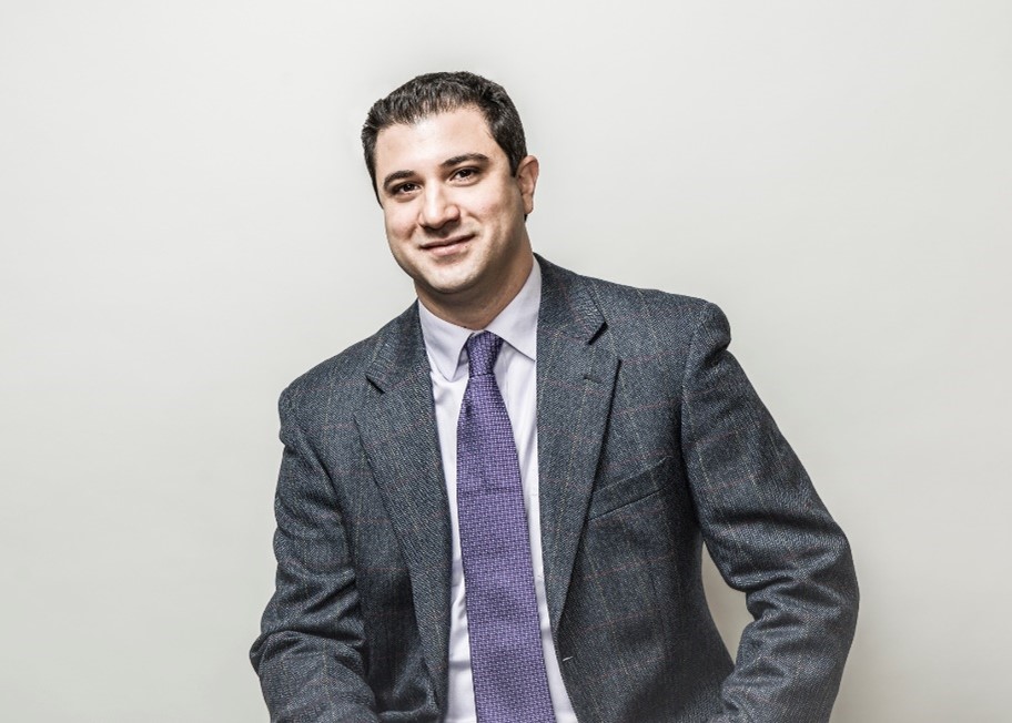
“When aiming to prevent heart attacks, a major point of interest is early detection of unstable atherosclerotic plaques,” says Nima Tabatabaei, associate professor in the Mechanical Engineering department at York University’s Lassonde School of Engineering.
These plaques are made up of fatty substances known as lipids, which can accumulate in different arteries throughout the body.
“If an atherosclerotic plaque ruptures, patients can experience a heart attack,” he says. “However, not all plaques will rupture. A major issue is that cardiologists don’t have a reliable way of distinguishing plaques that are problematic from those that are not.”
Over the past few years, Professor Tabatabaei, his former PhD student, Dr. Mohammad Hossein Salimi, and researcher at Massachusetts General Hospital and Harvard Medical School, Professor Martin Villiger, have been developing a new, intravascular imaging technology that can distinguish rupture-prone plaques from stable ones, based on their distinct structure and chemistry.
The explored method, Photo-Thermal Optical Coherence Tomography (PT-OCT), is an enhanced version of a biomedical imaging technique called Optical Coherence Tomography (OCT), which is primarily used to diagnose eye diseases.
PT-OCT utilizes two lasers as well as principles of interferometry to capture light scattering and absorption from biological tissues, thereby generating high-resolution structural images, co-registered with maps of tissue chemistry.
So, how is an eye imaging method a potential solution for heart disease?
“OCT is one of the most widely used optical imaging methods in clinical settings. Our idea was to include additional lasers to the system at specific wavelengths, allowing for absorption by the chemistry of unstable plaques,” says Professor Tabatabaei. “This permits the subsequent detection of distinct light absorption signatures. Our innovative method would support the production of co-registered images of both plaque structure and chemistry.”
In this way, cardiologists will have an objective tool for assessing the risk of atherosclerotic plaque rupture, allowing for early detection and prevention of heart attacks.
“Although PT-OCT is a promising method for imaging atherosclerotic plaques, it is currently not used in clinical settings for this purpose,” says Professor Tabatabaei. “Very few labs have access to this technology, and it can also be quite slow, complicated and hindered by noise.”
Throughout the past four years, Professor Tabatabaei and his research team have worked to address several fundamental and technological challenges of PT-OCT, while exploring innovative ways to improve this method for the purpose of imaging and characterizing atherosclerotic plaques.
In a recent paper, published in the prominent journal of Scientific Reports, part of Nature Publishing Portfolio, Professor Tabatabaei explored the use of an artificial intelligence (AI) method to improve the signal-to-noise ratio of PT-OCT signals and contrast of PT-OCT images. Neural networks were designed and trained with experimental data, allowing the system to better predict specific structural and chemical features in images. This work also created opportunities to significantly improve imaging speed and efficiency.
Through previous research, the team studied the implications of PT-OCT signals and their relationship with biological tissue, linking optical signals to corresponding chemical structures. They also investigated ways to improve additional qualities like accuracy, working toward practical use in clinical settings.
Detecting and characterizing atherosclerotic plaques, this imaging technology boasts the potential to transform the future of heart attack detection and treatment, ultimately improving patient outcomes. Moving forward, Professor Tabatabaei will continue to apply his expertise in biomedical optics to improve PT-OCT and explore additional applications, contributing to ongoing efforts of combatting heart disease, a leading cause of mortality in Canada and the United States.
Learn more about Professor Tabatabaei’s ongoing research efforts through his publications:
- Effects of lipid composition on Photothermal Optical Coherence Tomography signals
- Transient-mode Photothermal Optical Coherence Tomography
- Three-dimensional opto-thermo-mechanical model for predicting Photo-Thermal Optical Coherence Tomography responses in multilayer geometries
- Molecular-Specific Imaging of Tissue with Photo-Thermal Optical Coherence Tomography
- Artificial neural network for enhancing signal-to-noise ratio and contrast in Photothermal Optical Coherence Tomography